Match These Characteristics to the Appropriate Type of Microscope
Correctly label the following areas on a slide of neuronal tissue. A blood type also known as a blood group is a classification of blood based on the presence and absence of antibodies and inherited antigenic substances on the surface of red blood cells RBCs.

Introduction To The Microscope Ppt Video Online Download
Depending on the type of dye the positive or the negative ion may be the chromophore the colored ion.

. Match the type of muscle tissue with its description. Such a bias condition is called a forward bias. When on the other hand the bias condition is inversed which is called.
Coherent ultra 2 Ti. Stains or dyes contain salts made up of a positive ion and a negative ion. These strands are interrelated and are taught in an integrated way and in ways that are appropriate to specific local contexts.
Thus a large current flows across the junction. Place the characteristics with the appropriate muscular tissue. Focuses on the relationship between human behavior and criminal justice.
Depending on the type of dye the positive or the negative ion may be the chromophore the colored ion. Thanks for your kind help. Adiode solid state 405 nm bargon 458 nm 488 nm 514 nm cDPSS 561 nm dhelium neon 633 nm Class 4.
In addition to fixation staining is almost always applied to color certain features of a specimen before examining it under a light microscope. H-films are produced by stretching a sheet of polyvinyl alcohol to align the long-chain polymeric molecules which are subsequently impregnated with iodine. Light exiting the port in the microscope base is first passed through a neutral linear Polaroid HN-type polarizer to create plane-polarized light having a vibration vector that is confined to a single plane.
Stains or dyes contain salts made up of a positive ion and a negative ion. Sapphire 680-1080 nm Please advise which laser protective eyewear models will be appropriate for the above laser protection. Some specimens such as a drop of urine are already in a liquid form and can be deposited on the slide using a dropper.
A dissection and examination of the body. It is directly related to the angle of the cone which is formed between a point on the specimen and the front lens of the objective or condenser determined. Figured out a way to determine the type of blood from a dried bloodstain and began applying the new test to criminal investigations.
The Characteristics of a Fluorescence Microscope. Find an Affordable Stereo Microscope at New York Microscope Company. Sometimes the liquid used.
Rudi Rottenfusser in Methods in Cell Biology 2013. In Prep students learn about some Old Testament and New Testament stories that tell of a God of love the creator of all the goodness of Gods creation Gods special relationship with all of creation and Gods plan that people help each other to live. These antigens may be proteins carbohydrates glycoproteins or glycolipids depending on the blood group systemSome of these antigens are also present on the surface of other.
The simplest type of preparation is the wet mount in which the specimen is placed on the slide in a drop of liquid. The main parts of a fluorescent microscope overlap with the traditional light microscope. If you are looking to buy high-quality stereo microscopes we offer research grade common main objective microscopes which compete against the big four.
The other uncolored ion is called the counterion. The Numerical Aperture NA is the most important number associated with the light gathering ability of an objective or condenser. These modular stereo microscopes are based on the common main objective design.
However there are two main features that sets fluorescent microscope apart from the traditional microscope. Leica Nikon Olympus and Zeiss. One is the type of light source and the other is the use of specialized filter elements.
When a positive bias V is applied to the p-type region with respect to the n-type region the holes in the p-type region easily move into the n-type region and the electrons in the n-type region move in the opposite direction. Showed the uniqueness of fingerprints and how they could be used for identification. The other uncolored ion is called the counterion.
Solid specimens such as a skin scraping can be placed on the slide before adding a drop of liquid to prepare the wet mount. Match these cells found in connective tissues to their functions. Owns a confocal microscope which is equipped with the following lasers.
These films are less. In addition to fixation staining is almost always applied to color certain features of a specimen before examining it under a light microscope. Drag each label into the appropriate position to identify whether the statement depicts something true or false about the specimen.
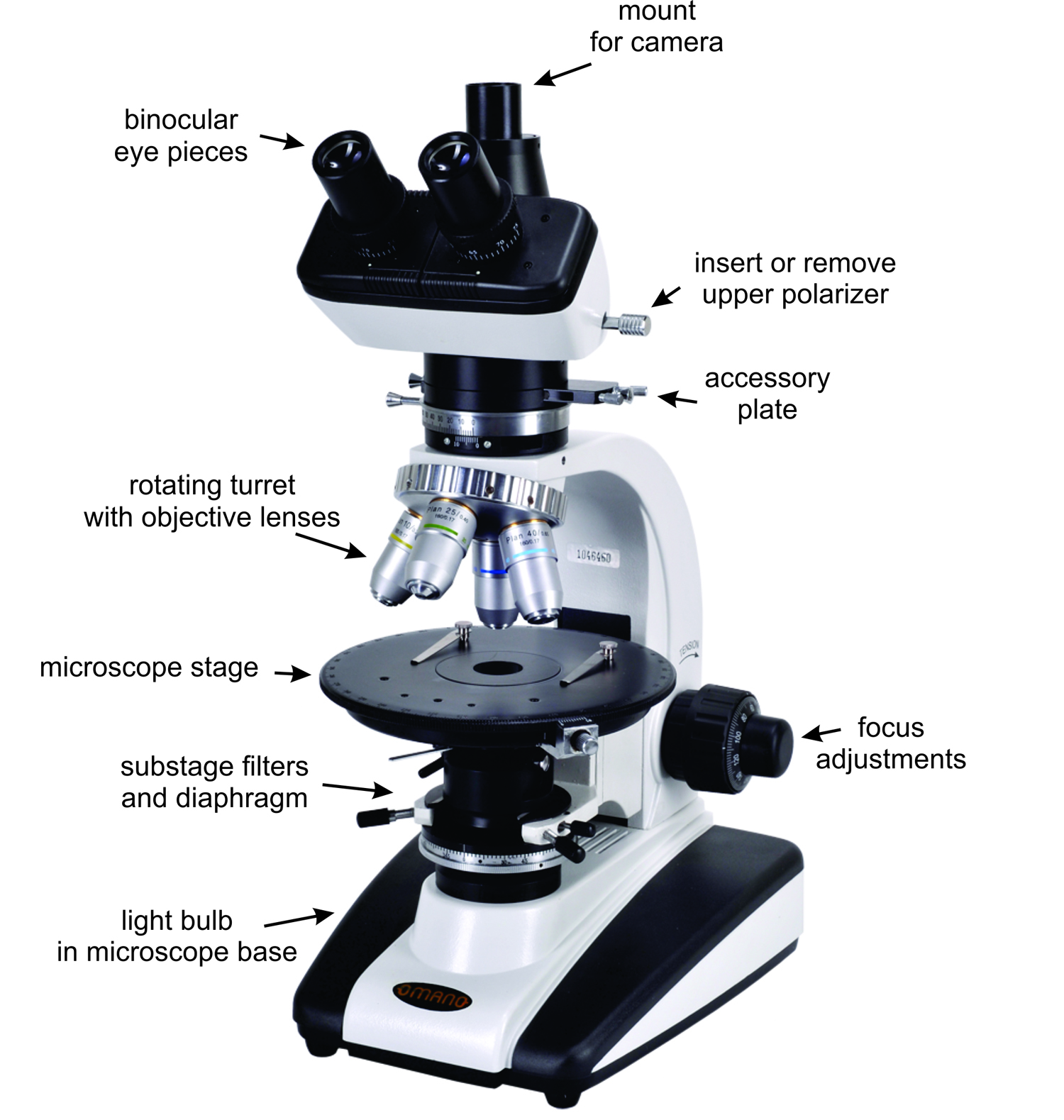
5 Optical Mineralogy Mineralogy

Mastering Microbiology Ch 3 Flashcards Quizlet
Mastering Microbiology Ch 3 Flashcards Quizlet
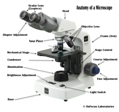
Microscope Parts And Functions

Comments
Post a Comment